心臓移植レシピエントカバーシート関連
心内膜心筋生検 Endomyocardial biopsy (EMB)
(例)半定量的評価:(–) (+) (++) (+++)
線維化
細胞浸潤
心筋細胞肥大
その他(コメント)
細胞浸潤
心筋細胞肥大
その他(コメント)
由谷親夫. 臨床医のための心筋生検アトラス (1997年3月) 医学書院 より
現在Amazon・紀伊國屋書店では入手できず。
p. 15 心筋細胞肥大度の基準(厚生省特発性心筋症調査研究班による診断マニュアル)
程度
右心室
左心室 (µm)
– 〜15 〜18
+ 16〜20 19〜23
++ 21〜25 24〜28
+++ 26〜 29〜
Cardiomyocytes of the right ventricle that are
≤15 μm in diameter are considered as not hypertrophic;
a diameter of up to 20 μm may indicate mild hypertrophy;
up to 25μm may indicate moderate hypertrophy;
between 25 and 30μm may be moderate to severe;
and a diameter >30μm is compatible with severe hypertrophy, based on our experience and unpublished data (Ishibashi-Ueda M et al. Circ J 2017;81:417-426)Significance and Value of Endomyocardial Biopsy Based on Our Own Experience
マイクロメーターを使用し、心筋細胞の横断面または縦断面で– 〜15 〜18
+ 16〜20 19〜23
++ 21〜25 24〜28
+++ 26〜 29〜
Cardiomyocytes of the right ventricle that are
≤15 μm in diameter are considered as not hypertrophic;
a diameter of up to 20 μm may indicate mild hypertrophy;
up to 25μm may indicate moderate hypertrophy;
between 25 and 30μm may be moderate to severe;
and a diameter >30μm is compatible with severe hypertrophy, based on our experience and unpublished data (Ishibashi-Ueda M et al. Circ J 2017;81:417-426)
核が存在する部位の短径を測定する。少なくとも30個の測定
が要求され、しかも心内膜直下の心筋細胞は計測しない。
核濃縮(核優勢)、心筋線維の異常分岐(非代償性高血圧)
文献: 岡田了三編: 心内膜心筋生検光学顕微鏡所見診断マニュアル. 厚生省特定疾患特発性心筋症調査研究班昭和53年度研究報告書, 1978
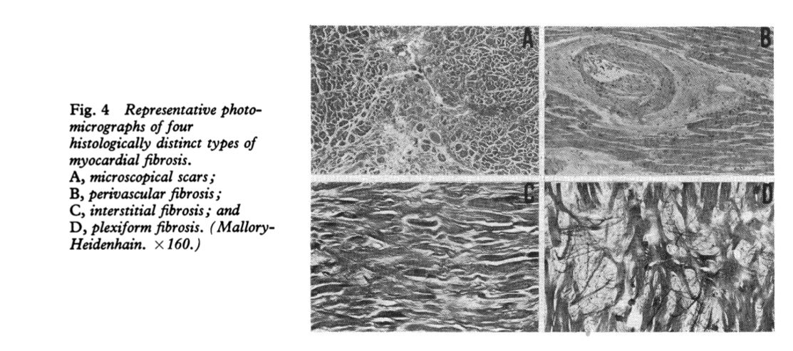
p.18 心筋線維化のgrading (staging?)
0: 心筋細胞を一重の線維が取り囲む
I: 二重異常に取り囲む (collagen fiberが二重,三重になる,写真では10%未満の線維化)
II: 心筋線維束を取り囲む(写真では心筋線維と同程度の幅の線維に取り囲まれている)
III: 上記の線維が癒合(写真では50%以上が線維化)
I: 二重異常に取り囲む (collagen fiberが二重,三重になる,写真では10%未満の線維化)
II: 心筋線維束を取り囲む(写真では心筋線維と同程度の幅の線維に取り囲まれている)
III: 上記の線維が癒合(写真では50%以上が線維化)
文献: Yutani C, Go S, Kamiya T, et al. Cardiac biopsy of Kawasaki disease. Arch Pathol Lab Med. 1981 Sep;105(9):470-3.
線維化の種類
interstitial (高血圧), replacement(心筋梗塞), perivascular (膠原病), plexiform (錯綜配列)

心内膜肥厚 endocardial fibrous thickening or endocardial fibroelastosis:
elastica van-Gieson staining is needed.
The presence of ≥10 elastic fiber layers in endocardial fibrous thickening is considered abnormal proliferation.
p. 22 細胞浸潤と心筋炎
5/hpf以上を有意な炎症細胞浸潤と見なす(Edwards) (種類: ly,
mono, eos)
軽度(散在性, perivascular, 1Rではone focusまでの心筋傷害-細胞質の淡明化・核優勢・scalloping)
中等度(2Rでは2つ以上の心筋傷害)
高度(3Rではびまん性, 多数の心筋傷害; 浮腫/出血/血管炎があってもなくても)
軽度(散在性, perivascular, 1Rではone focusまでの心筋傷害-細胞質の淡明化・核優勢・scalloping)
中等度(2Rでは2つ以上の心筋傷害)
高度(3Rではびまん性, 多数の心筋傷害; 浮腫/出血/血管炎があってもなくても)
文献: Edwards WD, Holmes DR Jr, Reeder GS. Diagnosis of active lymphocytic myocarditis
by endomyocardial biopsy: quantitative criteria for light microscopy. Mayo Clin Proc. 1982 Jul;57(7):419-25.
Stewart S, Winters GL, Fishbein MC, et al. Revision of the 1990 Working Formulation for
the Standardization of Nomenclature in the Diagnosis of Heart Rejection J Heart Lung Transplant 24:1710-20, 2005.
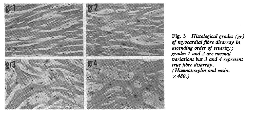
p. 33 心筋錯綜配列grading
0
1 focal, mild
2 focal, distinct
3 multiple - plexiform, 核異常
1 focal, mild
2 focal, distinct
3 multiple - plexiform, 核異常
文献: St John Sutton MG, Lie JT, Anderson KR, et al: Histopathological specificity of
hypertrophic obstructive cardiomyopathy. Myocardial fibre disarray and myocardial fibrosis. Br Heart J. 1980 Oct;44(4):433-43.
