1

#1 Boston–Kyoto Surgical Pathology Update 2013

Slide Seminar Thumbnails

originally http://www2.massgeneral.org/pathology/hms/Kyoto13.htm (deleted)

  
WF_04

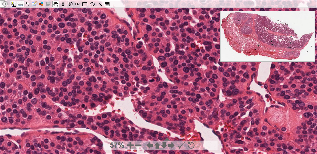
69 year-old female with 5 cm left thyroid mass.

Insular variant of poorly differentiated thyroid carcinoma

William C Faquin M.D.,PhD
WF_06

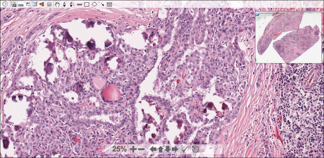
6 year-old female with lymphadenopathy and bilateral thyroid masses.

Diffuse sclerosing variant of papillary thyroid carcinoma
William C Faquin M.D.,PhD  
NC_01

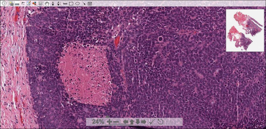
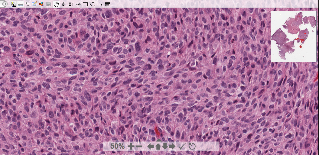
70 year old female with 5 cm mass involving right pyriform sinus and larynx

Basaloid squamous cell carcinoma

William C Faquin M.D.,PhD
Day1_case 4
Orbital tumor.

Subconjunctival herniated orbital fat
G.Petur Nielsen M.D
footmass
Foot mass

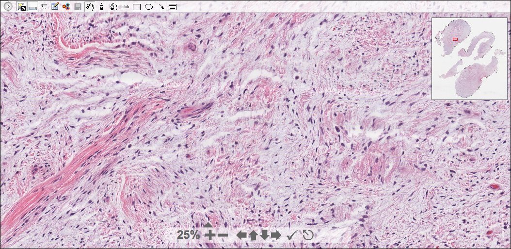
Hemosiderotic fibrolipomatous tumor
G.Petur Nielsen M.D
GUN_01.jpg
What is the location ?

Finger
(Superficial acral fibromyxoma)
G.Petur Nielsen M.D
images/Day1_case1.jpg
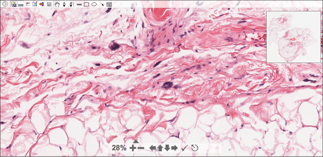
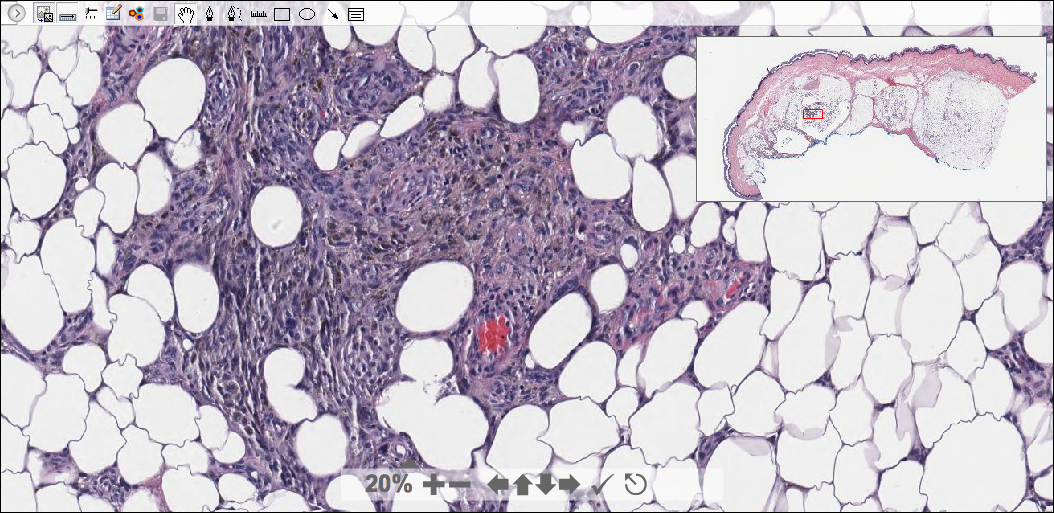
Retroperitoneal mass.

Sclerosing extramedullary hematopoietic tumor
G.Petur Nielsen M.D  
images/Day1f.jpg Soft tissue tumor

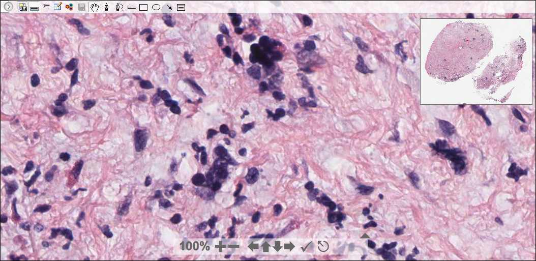
Dedifferentiated metastatic malignant melanoma
G.Petur Nielsen M.D  

 1
Insular/trabecular patterns, occasional mitoses, nuclear features different from typical papillary carcinoma.

2
Numerous psammoma bodies and morule formation, in Hashimoto thyroiditis-like dense lymphocytic and sclerotic background

3
Comedo necrosis and numerous mitoses in basaloid cells

4
The lesion contains floret cells that may mimic pleomorphic lipoma and atypical lipomatous tumor.

5
A hemosiderotic fibrohistiocytic lipomatous lesion, or hemosiderotic fibrolipomatous tumor.
Consistent t(1;10) with rearrangements of TGFBR3 and MGEA5 in both myxoinflammatory fibroblastic sarcoma and hemosiderotic fibrolipomatous tumor.

6
Bland spindle cells with CD34 and EMA positivity

7
Large cells were Megakryocytic cells, as seen in original myeloproliferative disorders.

8
All melanoma markers including S-100 were negative ("dedifferentiated"). BRAF+ as was previous malignant melanoma of the skin.